
Pin on A&P.4.Heart.Lung
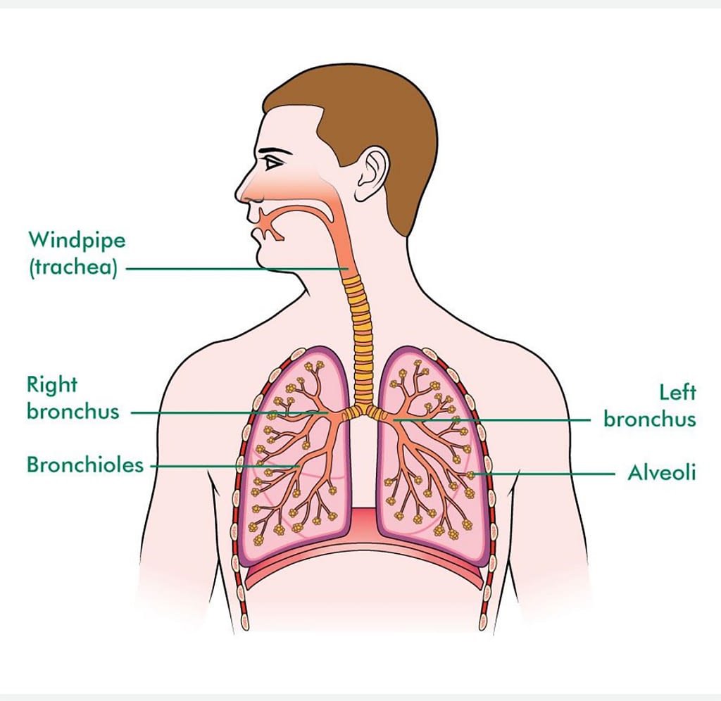
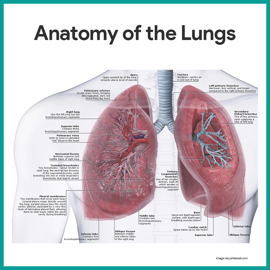
The purpose of the lung is to provide oxygen to the blood. The respiratory system divides into airways and lung parenchyma. The airways consist of the bronchus, which bifurcates off the trachea and divides into bronchioles and then further into alveoli. The parenchyma is responsible for gas exchange and includes the alveoli, alveolar ducts, and bronchioles. Lungs have a spongy texture and have.
A healthy lung has a pinkish appearance, and if you could see it outside the body, it would look
The respiratory system, also called the pulmonary system, consists of several organs that function as a whole to oxygenate the body through the process of respiration (breathing). This process involves inhaling air and conducting it to the lungs where gas exchange occurs, in which oxygen is extracted from the air, and carbon dioxide expelled.
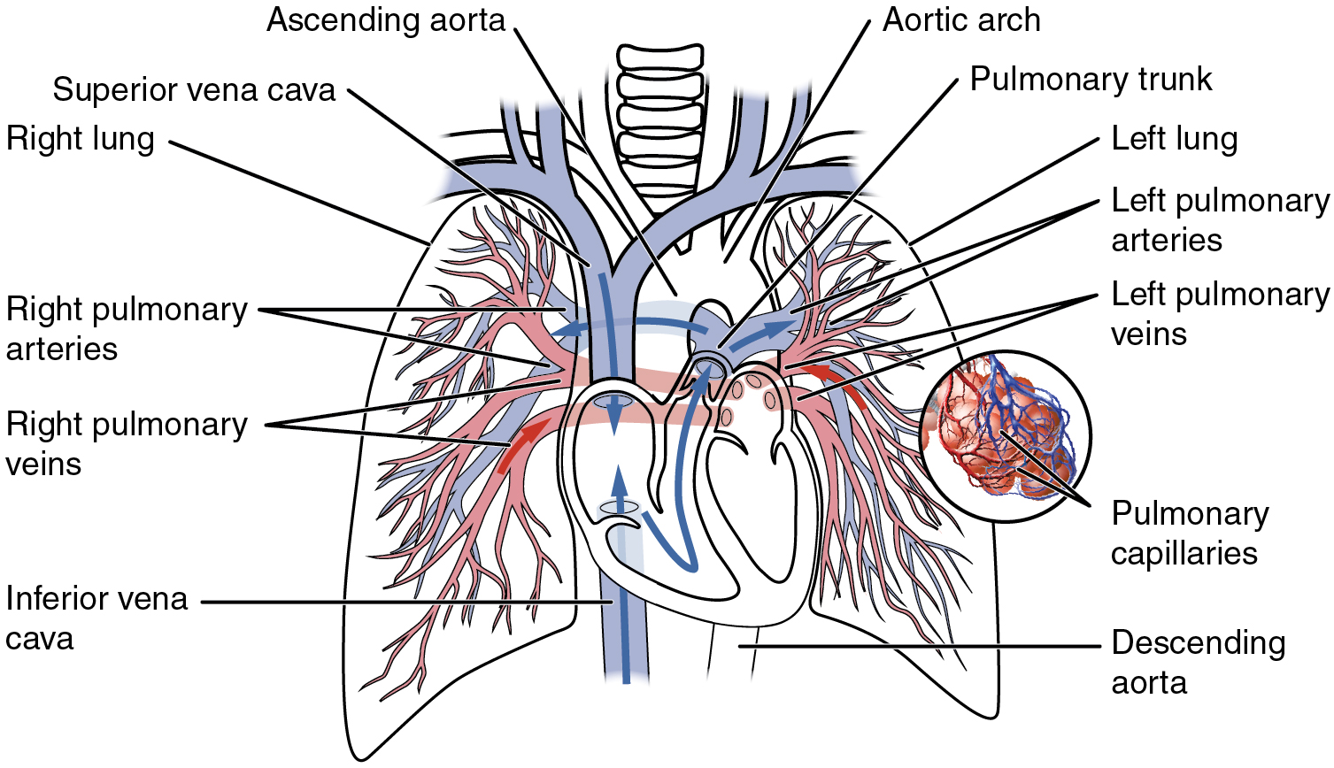
Circulatory Pathways · Anatomy and Physiology
This chart of the RESPIRATORY SYSTEM shows how you breathe. Breathing is the process that brings oxygen in the air into your lungs and moves oxygen and through your body. Our lungs remove the oxygen and pass it through our bloodstream, where it's carried off to the tissues and organs that allow us to walk, talk, and move.Our lungs also take carbon dioxide from our blood and release it into the.
Lungs 101 Journey to Health with Priya
Lungs are a pair of respiratory organs situated in a thoracic cavity. Right and left lung are separated by the mediastinum. Texture -- Spongy. Color - Young - brown. Adults -- mottled black due to deposition of carbon particles. Weight-. Right lung - 600 gms. Left lung - 550 gms.
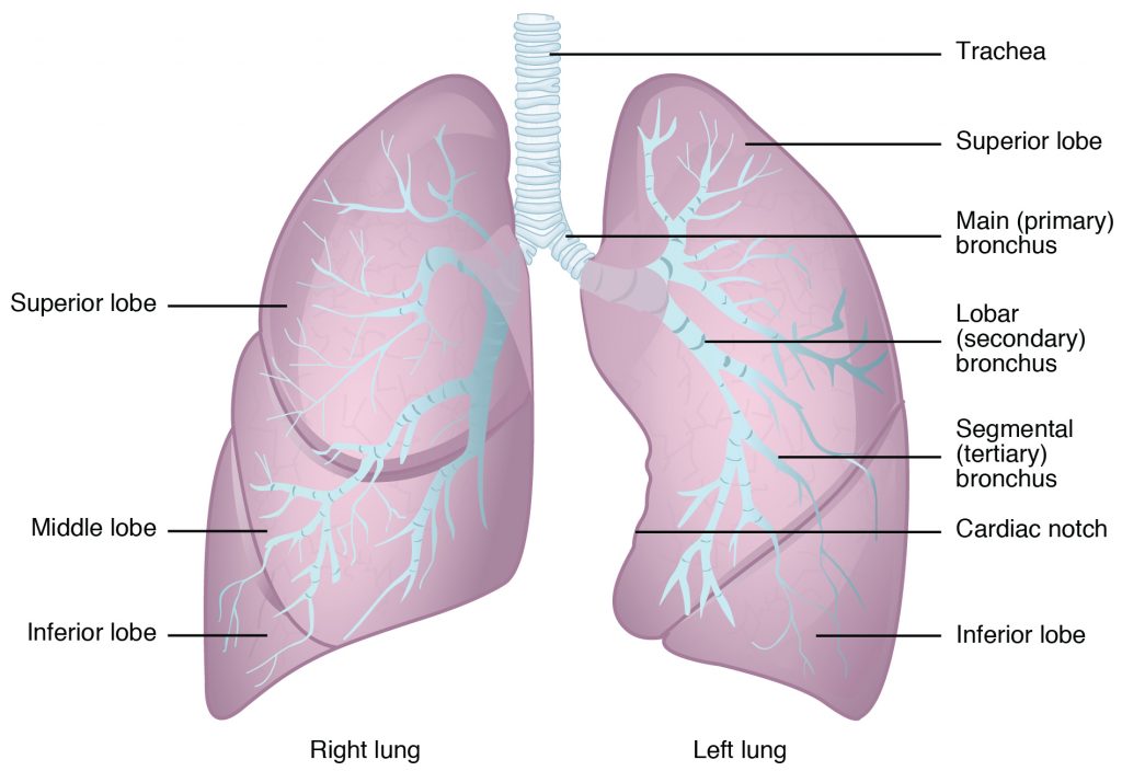
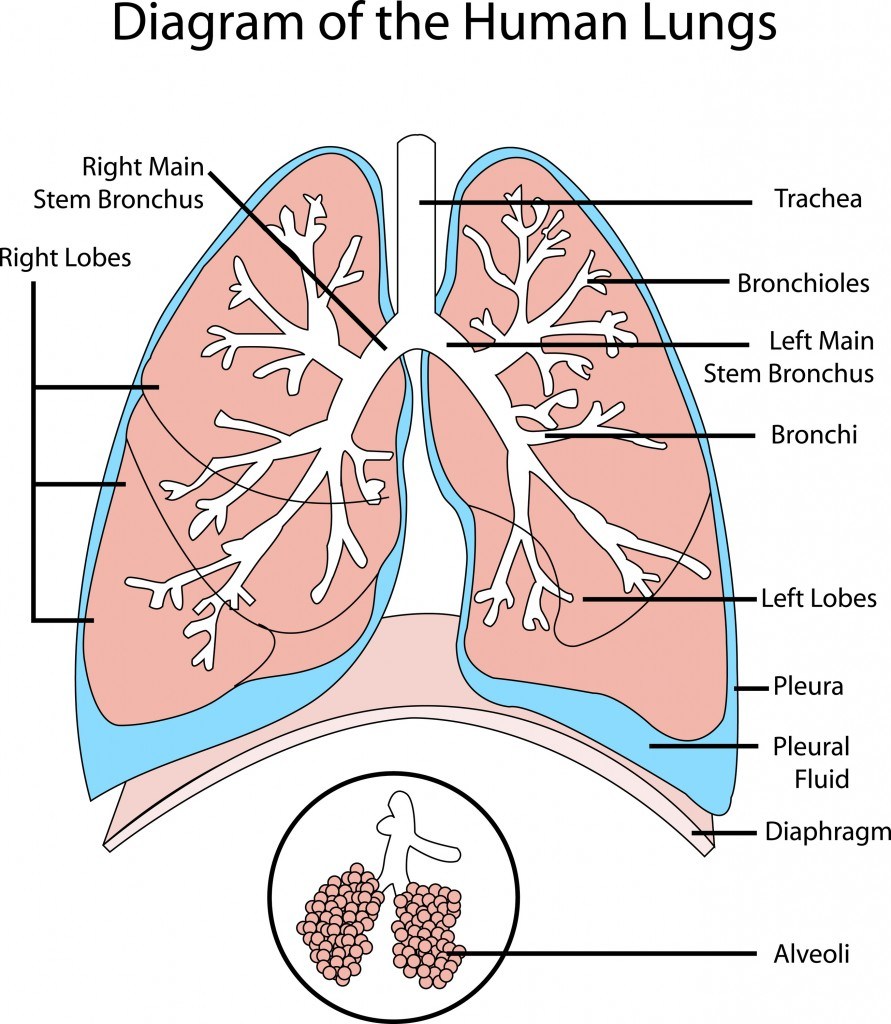
Diagram Of The Lungs With Labels Labeling Of The Lungs Label The Lungs Diagram Diagram Of Lungs
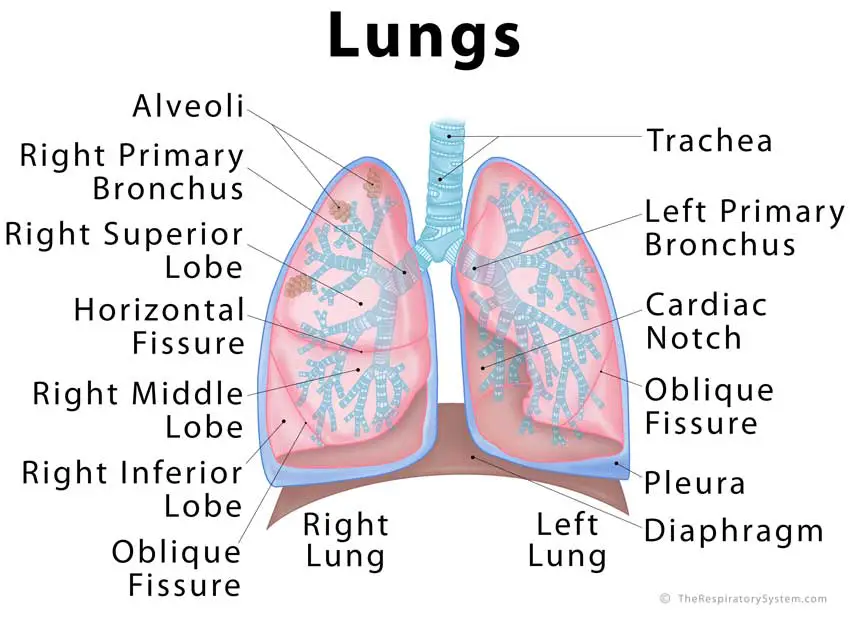
The lung on your right side is divided into three lobes: the superior, the middle and the inferior. It's shorter than your left lung, but also wider than your left lung. Both of your lungs are covered with a protective covering called pleural tissue. Left lung. Your left lung has two lobes: the superior and the interior.

Lung Structure BioNinja
The respiratory system aids the body in the exchange of gases between the air and blood, and between the blood and the body's billions of cells. It includes air passages, pulmonary vessels, the.
Respiratory System Anatomy and Physiology Nurseslabs
The lungs also get rid of carbon dioxide, a waste product of the cells. The lungs are a pair of cone-shaped organs made up of spongy, pinkish-gray tissue. They take up most of the space in the chest (thorax). The lungs are surrounded by a membrane (pleura). The lungs are separated from each other by the mediastinum, an area that contains the:
Auscultation how to do chest, lung and heart auscultation
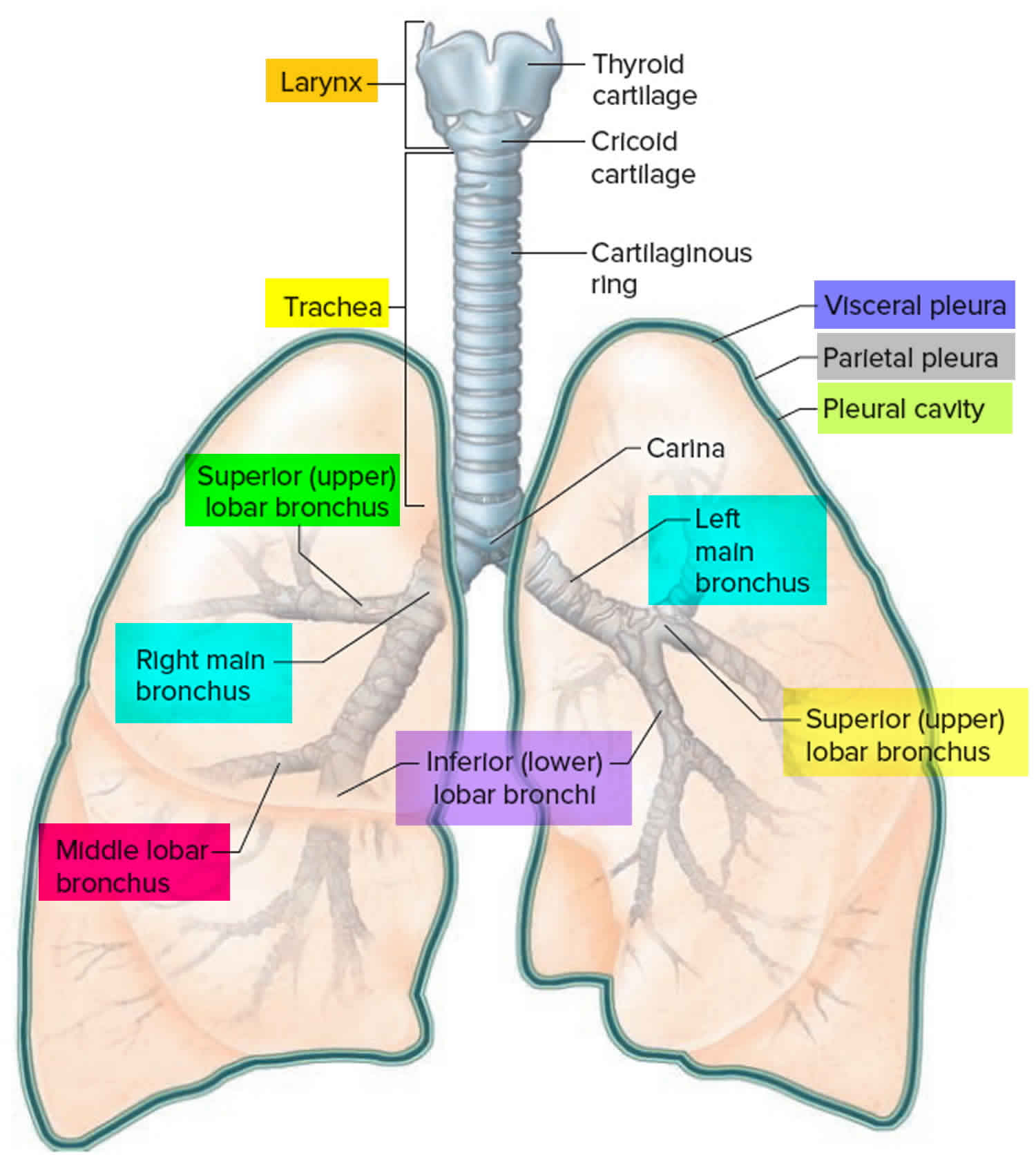
Respiratory system labeled Take a look at the labeled diagram of the respiratory system above. As you can see, there are several structures to learn. Spend a few minutes reviewing the name and location of each one, then try testing your knowledge by filling in your own diagram of the respiratory system (unlabeled) using the PDF download below.
Lung Diagram With Labels General Wiring Diagram
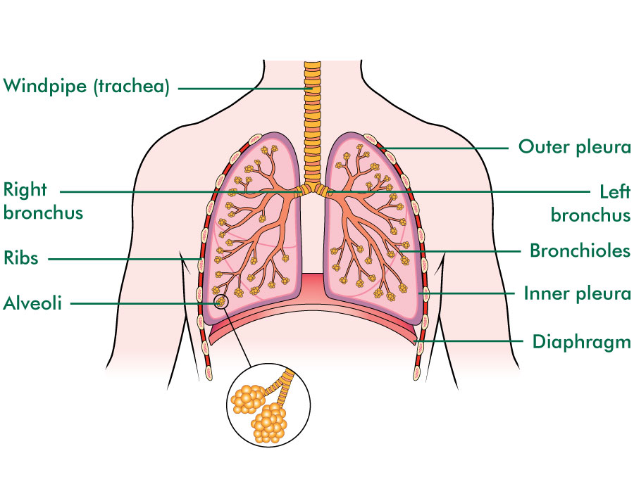
The lungs are respiratory organs - we use them to breathe. We breathe in order to get oxygen (O 2) and to get rid of carbon dioxide (CO 2 ). We breathe in through the nose or mouth. The air then goes through the larynx and the trachea (also called the windpipe) and into the lungs. We breathe by using the diaphragm, a muscular membrane under.

Lung Diagrams 2017 101 Diagrams
Where are the Lungs Located. The lungs are located a little toward the posterior part of the human body, just below the collarbone, extending down to the diaphragm, the muscular partition that separates the chest and abdominal cavities.The left and right lungs are situated on the two sides of the body with the heart, another vital organ in the thoracic cavity, located a little in front of, and.

Lung Diagrams Diagram Link
bronchial tree: the system of airways within the lungs, which bring air from the trachea to the lung's tiny air sacs (alveoli). cardiac notch: the indentation in the left lung that provides room for the heart. diaphragm: a muscular membrane under the lungs. larynx: a muscular structure at the top of the trachea, containing the vocal cords.

Medical Education Chart of Biology for Lungs Diagram. Vector illustration Ad , sponsored,
The lungs are a major organ that is part of the respiratory system, taking in fresh air and getting rid of old, stale air. This mechanism of breathing also helps to allow you to talk. By taking in fresh air, the lungs are able to help oxygenate blood to be carried around your body. This is done by inhaling the air and bringing it in toward the.

Labeled diagram of the lungs/respiratory system.
View Original Image at Full Size. Labeled diagram of the lungs/respiratory system. Image 37789 is a 1125 by 1408 pixel WebP Uploaded: Jan10 14. Last Modified: 2014-01-10 12:15:34
Anatomy of LUNGS and MCQs for NEET, GPAT,Nursing Exams Gpatindia Pharmacy Jobs, Admissions
Respiratory system - Edexcel Respiratory system structure and function. The respiratory system transports oxygen from the air we breathe, through a system of tubes, into our lungs and then.
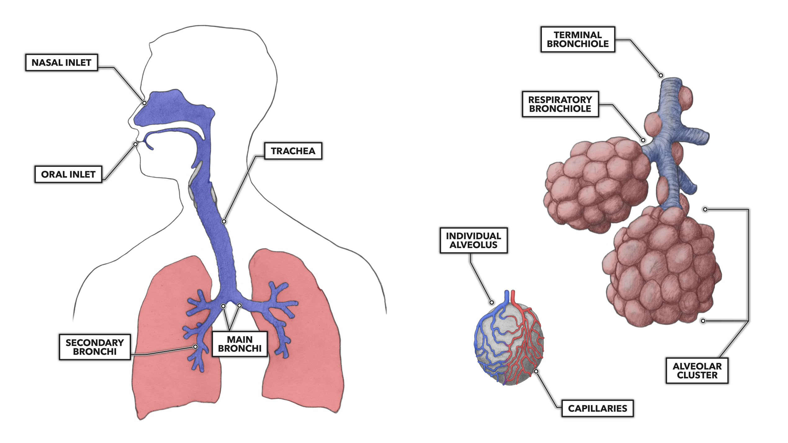
CrossFit Lung Anatomy The Airway and Alveoli
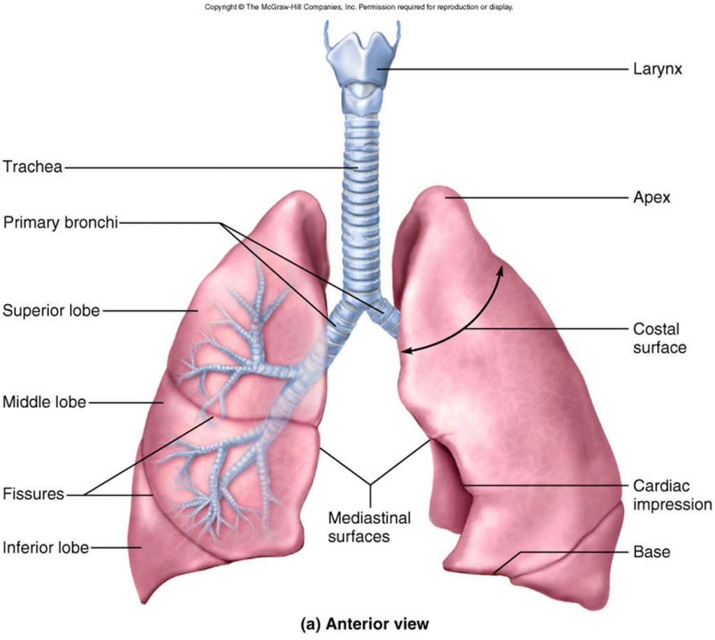
Ease into this topic, simplify your learning and improve your retention using Kenhub's respiratory system quizzes and labeled diagrams! Lobes of the right lung The superior lobe of the right lung is located above the horizontal fissure and anterosuperior to the oblique fissure. It contains the apex, majority of the upper part of the costal.
Lungs clipart labeled, Lungs labeled Transparent FREE for download on WebStockReview 2023
The costal surface of the lung borders the ribs. The mediastinal surface faces the midline. Figure 22.2.1 Gross Anatomy of the Lungs. Each lung is composed of smaller units called lobes. Fissures separate these lobes from each other. The right lung consists of three lobes: the superior, middle, and inferior lobes.